CRUK City of London Centre 2024 Image Competition Winners
Many thanks to everyone who submitted their images to the CRUK City of London competition, and congratulations to the winning entrants.
First prize
Mabel Baxter Dalrymple
King’s College London
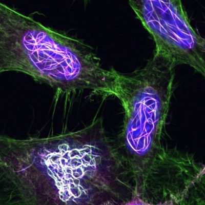
Knot Actin As You Know It
Mabel Baxter Dalrymple’s PhD is funded by the Cancer Research UK City of London Centre, and focuses on examining filamentous actin in the nuclei of breast cancer cells.
Actin, a structural protein, is a well-studied ‘housekeeping gene’ that is present in all cells and is essential for maintaining basic function. Actin assembles dynamic and diverse structures contributing to the cell skeleton. This enables cells to move, divide, grow, and sense their surrounding environment.
Here, fluorescence microscopy is used to reveal filamentous actin in and around the nuclei (in blue) of single breast cancer cells, making intricate knot-like structures (green, grey and magenta).
Mabel Baxter Dalrymple was interested to study what happens when actin is stabilised in its polymerised state as a filament of multiple molecules (known as ‘F-actin’) by using genetically mutated actin variant inserted into breast cancer cells and targeted to the nucleus.
Using breast cancer tissue samples, she has found that there is more nuclear filamentous actin in more advanced breast cancer, suggesting that it has a role in supporting the progression of cancer through regulating the cancer genome and cell invasiveness.
Ultimately, this work could inform our understanding of breast cancer development and prognosis, supporting decisions around treatment.
Joint second prize
Panoraia Kotantaki
Barts Cancer Institute/QMUL
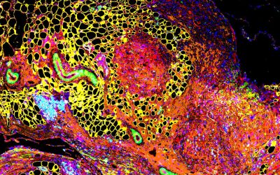
Breaking the barriers, unleashing anti-tumour troops in the metastatic niche
Dr Kotantaki’s image was captured during her research into the use of chemotherapy drugs in a novel combination with a lysyl oxidase inhibitor.
Dr Kotantaki and her colleague Dr Florian Laforêts are investigating how this approach shrinks metastatic (secondary) ovarian cancer tumours that are growing in the omentum, a lining of fatty adipose tissue in the abdomen.
The image shows adipocytes – normal fat cells – in yellow, reclaiming their space as the peri-tumoral fibrotic barrier (in orange) collapses and the tumour (in red) shrinks. In light blue, we see CD8+ cytotoxic T cells, immune cells that have been recruited to fight the tumour.
Metastatic ovarian tumours are typically highly fibrous, making them harder to treat or remove. Dr Kotantaki’s research offers hope that this new combination of treatments could be used to shrink them. In further good news for patients, the new lysyl oxidase inhibitor can be taken orally, and Dr Kotantaki’s studies in mice show excellent drug tolerability, meaning fewer unpleasant side effects.
To capture this image, Dr Kotantaki used the City of London Centre’s Cell DIVE Imaging Platform, based at the Blizard Institute and worked in collaboration with Joseph Hartlebury and Dr Florian Laforêts.
Joint second prize
Irina Titkova
Barts Cancer Institute/QMUL
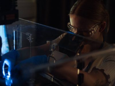
After 5pm on Friday
Dr Titkova’s research focuses on ovarian cancer, targeting a new pathway with drug combinations to try to induce synthetic lethality.
Many stunning microscopic images were submitted to our competition, but Dr Titkova’s stood out as it shows the other side of the lens. A professional photographer, she had planned to produce a portrait of her entire lab group, but this was not possible to orchestrate on a busy afternoon.
Instead, her winning image was spontaneous. It captures her colleague Caroline’s intense concentration as she powered through her work on a Friday afternoon in the final year of her PhD investigating colorectal adenocarcinoma. As the microscope glows in a dimly lit laboratory, Caroline’s gaze is fixed on the microscopic world, and the viewer is invited to contemplate what discoveries may await.
The image is a tribute to the focus and dedication required for a career in research science.
In her spare time, Dr Titkova works as a science communicator and graphic designer, capturing the beauty of science. She took this spectacular image using a Panasonic Lumix GH5.
Joint third prize
Sarah Duncan
Barts Cancer Institute/QMUL
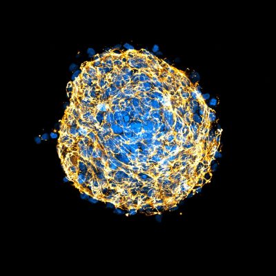
Extracellular matrix deposition in a pancreatic cancer spheroid
Dr Duncan’s research focuses on pancreatic cancer. It is thought that up to 90% of the volume of a pancreatic tumour is made of connective tissue that is non-cancerous. This makes it especially difficult to treat with cancer-targeting drugs. Dr Duncan aims to further our understanding of what causes this tissue to develop.
The proteins in connective tissue are deposited by pancreatic stellate cells, star-shaped fibroblasts that are inactive in healthy tissue. Cancer cells can cause the stellate cells to become activated and lead to increased deposition of non-cancerous connective tissue.
Previous attempts to quantify this phenomenon using 2D models had been unsuccessful, so Dr Duncan created a 3D spheroid model. Immunofluorescent staining enabled the visualisation of protein deposits using a point scanning confocal microscope, which uses a laser to create high resolution images.
Dr Duncan’s image shows pancreatic cancer cells and pancreatic stellate cells in blue, and the extracellular matrix protein, fibronectin, in yellow.
Dr Duncan aims to gather information about the role played in the activation of stellate cells by extracellular vesicles, key mediators of cell-cell communication. This will support the development of a treatment strategy to reduce the activation of stellate cells by cancer cells and decrease the deposition of fibrous tissue in pancreatic cancer tumours, hopefully making them more responsive to treatment.
Joint third prize
Florian Laforêts
Barts Cancer Institute/QMUL
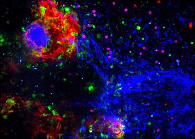
Live immune cells attacking cancer cells in a human tumour
Dr Laforêts’s image is a still from a video clip, produced while investigating the movement of cells within live human tissue. It shows an ovarian cancer tumour.
In pink, we see the cytotoxic lymphocytes, cells that aim to kill infected or malignant cells in the body. Here, they move within the tumour to try to kill cancer cells, shown in green. They are often impeded in their movements by other cells, such as macrophages (shown in red) and fibres of the extracellular matrix, shown in blue.
This image captures the way a tumour represses the movement and anti-cancer activity of some immune cells. This reduces chemotherapy efficiency and is one of the reasons why fewer than 35% of ovarian cancer patients survive beyond 5 years post diagnosis. Therefore, one of the challenges of Dr Laforêts’s research is to boost the immune system to enhance chemotherapy and improve survival.
Dr Laforêts submitted this image to show how science and art coexist and how, together, they can serve a common goal: reaching out to a wider audience and reminding us why research is so important.
