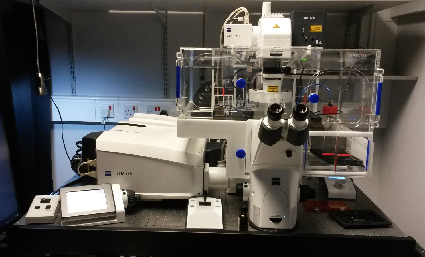
Zeiss LSM 880 Confocal Microscope with Airyscan Fast
The Zeiss LSM 880 Confocal Microscope with Airyscan Fast is located in the Light Microscopy Core Facility at CRUK Barts Centre.
The Microscopy Service provides access to imaging equipment suiting a wide range of applications. Equipment includes advanced confocal microscope systems, laser microdissection, various time lapse options and scanners for digital pathology.

Contact
Equipment
The LSM 880 point-scanning confocal microscope is inverted and fitted with an environmental control chamber, allowing users to perform live imaging experiments that can be recorded as a time series. The scanning stage has inserts to accommodate slides, dishes and plates. The microscope is equipped with a Fast Z piezo insert for fast acquisition of Z stacks and Definite Focus 2 for keeping focus during time series. The system includes an Airyscan super resolution detector and Fast module.
Axio Observer Z1 motorised stand with 3 position optovar turret for DAPI, GFP and Cy3.
Available objectives are: Plan-Apochromat 63x/1.40 Oil DIC; Plan-Apochromat 40x/1.30 Oil DIC; PA po 20x/0.80; PA po 10x/0.45 and LD 40x/1.30 Corr.
Lasers: Diode 405nm, multi-line Argon 457/ 488/ 514 nm, HeNe 561nm and HeNe 633nm
The 34 channel spectral system comprises two PMT and 32 x channel GaAsP detectors.
Zen software modules for Z-stacking, Tiling, Time Series, Bleaching, Positions, Regions and FRAP.
Bookings can be made using iLab, an online system for managing Queen Mary’s research laboratories and equipment – https://www.qmul.ac.uk/ilab/
The price for external users is £40 per hour.
Training for the LSM 880 with Airyscan Fast is compulsory and free of charge. Training can be requested through iLab or by emailing l.j.hammond@qmul.ac.uk.
The facility provide advice for experimental and staining protocols, in addition to help for setting up appropriate imaging configurations.

